ESC
Molecular Biology

Genetics & Molecular Biology

I apply a comprehensive range of genetic and molecular techniques — from CRISPR editing and RNA interference to molecular cloning and viral transduction — to probe protein trafficking, organelle stress, and cellular quality control.

CRISPR/Cas9 siRNA / shRNA / ASO / SSO Molecular Cloning Lentiviral / AAV / Adenoviral IP / Fractionation Lysosomal Biology qPCR Data Flow Cytometry Data Virology Western Blot / ELISA / MSD PCR / qPCR / ddPCR
01

CRISPR & Gene Perturbation

I have generated multiple CRISPR-edited iPSC cell lines (Parkin KO, α-synuclein KO, LRRK2 KO, GBA KO) to assess protein function and mimic genetic forms of neurodegenerative diseases. GBA knockout heightened vulnerability to fibril-induced inclusions, whereas α-synuclein knockout abolished pathology entirely. Validation includes PCR, Western blotting, karyotyping, and ICC for pluripotency markers.

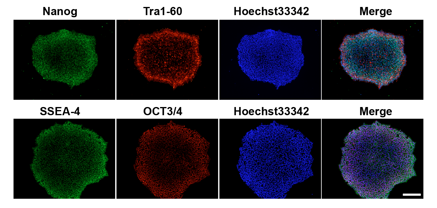
iPSC Colony QC & Pluripotency

I maintain rigorous QC across all iPSC lines including morphology assessment, pluripotency marker ICC (Oct4, Sox2, Nanog, TRA-1-60), mycoplasma testing, and karyotype validation. Colony morphology is monitored throughout expansion to ensure undifferentiated state.

iPSC colony QC: phase contrast morphology, pluripotency ICC staining panels
Quality control imaging of iPSC colonies showing pluripotency marker expression, morphology, and karyotype confirmation.
02

RNA Interference & Oligonucleotide Biology

I design, optimize, and validate siRNA, shRNA, ASO, and SSO-mediated knockdown assays in iPSC-derived neurons and cancer cell lines.

Fig. — CHC Knockdown: shRNA vs siRNA Comparison
Clathrin heavy chain (CHC) mRNA remaining after knockdown by shRNA and siRNA in U2OS cells
RT-qPCR normalized to GAPDH. 72h post-transfection (siRNA) or 7d post-transduction (shRNA). n=3 biological replicates.
Fig. — ASO/SSO-Mediated α-Synuclein Modulation
SNCA mRNA and protein levels in iPSC-derived DA neurons after ASO/SSO treatment (dose-response)
Gymnotic delivery, 7-day treatment. RT-qPCR (mRNA) and Western blot densitometry (protein) normalized to scrambled control.
Delivery methods include gymnotic uptake (media addition), lipid-based transfection, and electroporation/nucleofection. I integrate RT-qPCR, protein-level (Western/ELISA), and functional neuronal readouts to assess potency, durability, specificity, and mechanism-of-action.

I have miniaturized assays into 96/384-well formats with SOP and repeatability metrics for scalable, screening-ready execution, translating RNAi results into clear go/no-go and target-prioritization recommendations for preclinical programs.

03

Molecular Cloning & Construct Design

End-to-end cloning workflows including restriction cloning, Gibson assembly, Golden Gate cloning, PCR/primer design, bacterial transformation, colony screening, plasmid preparation (MiniPrep/MaxiPrep), Sanger sequencing, and endotoxin-free DNA prep. I design constructs for membrane-associated and endolysosomal proteins, with expression verification by flow cytometry and/or immunoassay. Stable cell line generation uses selection markers (Puromycin, G418, Blasticidin) with single-cell cloning and pool evaluation.

04

Viral Transduction & Expression

I produce and optimize lentiviral, adenoviral, and AAV/rAAV constructs for transduction in iPSC-derived neurons and mammalian cell lines. Key applications include: (1) lentiviral shRNA knockdown of LAMP2, which I demonstrated is sufficient to drive α-synuclein inclusion formation even without immune challenge — a critical finding from my Nature Neuroscience 2024 paper; (2) adenoviral overexpression of α-synuclein-HA to show that endogenous protein is actively recruited into pathological inclusions; and (3) reporter construct expression for live-cell tracking. I handle full vector design (cassettes, promoters, tags), MOI optimization, titering, and biosafety compliance across BSL-2 workflows.

LAMP2 shRNA knockdown: Western blot validation and confocal showing inclusion formation
Confocal immunohistochemistry showing successful LAMP2 shRNA knockdown (reduced LAMP2 signal) in iPSC-derived dopaminergic neurons.

Lentiviral shRNA-mediated LAMP2 knockdown in iPSC-derived DA neurons. Western blot confirms >80% reduction in LAMP2 protein. Confocal shows that LAMP2 loss alone — without immune challenge — is sufficient to induce α-synuclein inclusion formation, establishing lysosomal dysfunction as the critical vulnerability in Lewy body pathogenesis. From Bayati et al., Nature Neuroscience 2024.

Adenovirus α-synuclein-HA overexpression with pSyn panels and Western blot across treatment conditions
Confocal time-course showing adenoviral α-synuclein-HA overexpression in iPSC neurons ± IFN-γ treatment, revealing IFN-induced accumulation.

Adenoviral overexpression of α-synuclein-HA in iPSC-derived DA neurons. Phospho-synuclein (pSyn S129) accumulation is dramatically enhanced under dual-hit conditions (PFF + IFN-γ), confirming that endogenous α-synuclein is recruited into pathological inclusions. Western blot quantification shows dose-dependent pSyn increase. Bayati et al., Nature Neuroscience 2024.

05

Lysosomal Biology & Protein Biochemistry

My work on lysosomal biology spans from organelle-level mechanistic studies to biochemical characterization. I showed that IFN-γ specifically downregulates LAMP1, LAMP2, TFEB, and NRF2 in DA neurons — but not cortical neurons — creating a selective vulnerability window for Lewy body formation (Nature Neuroscience 2024). I use galectin-3 as a reporter for lysosomal membrane permeabilization, revealing that fibrils rupture lysosomes and escape into the cytoplasm. My Lyso-IP approach (HA-tagged TMEM192-RFP + magnetic bead immunoprecipitation) confirmed PFF arrival at lysosomes within 2 minutes of uptake.

For protein biochemistry, I perform recombinant α-synuclein purification (Ni-NTA affinity), preformed fibril (PFF) generation (37°C shaking, 5 days, sonication), size exclusion chromatography (SEC), and DLS particle sizing. I developed a custom nanogold-PFF conjugation protocol — published in STAR Protocols (2023) as first author — enabling direct EM visualization of fibril trafficking without relying on antibody specificity.

Fig. — SEC: α-Synuclein Purification Profile
Size exclusion chromatography of recombinant α-synuclein — monomer, oligomer, and fibril fractions
Superdex 200 10/300 GL column. UV absorbance at 280 nm.
Fig. — DLS: Particle Size Distribution
Dynamic light scattering of α-synuclein monomer vs fibril preparations
Z-average diameter. Monomer ~3 nm, fibrils ~180 nm with polydispersity.
06

Immunoprecipitation & Biochemical Fractionation

I apply biochemical fractionation and immunoprecipitation (IP/Co-IP) techniques to resolve protein compartmentalization and interaction networks. Key applications include: (1) sequential detergent extraction (Triton-soluble → SDS-soluble → insoluble pellet) to separate soluble, membrane-associated, and aggregated protein pools; (2) Lyso-IP (HA-tagged TMEM192-RFP + magnetic bead immunoprecipitation) to isolate intact lysosomes and confirm fibril cargo arrival within 2 minutes; and (3) native co-IP workflows for capturing transient protein–protein interactions under physiological conditions. Downstream readouts include Western blot, mass spectrometry, and activity assays.

Subcellular fractionation Western blots: HDAC6 and α-synuclein from sequential extraction
Western blot of biochemical fractionation (soluble, membrane, and pellet) showing α-synuclein distribution with and without inclusion formation.

Biochemical fractionation of α-synuclein inclusions from iPSC-derived DA neurons. Sequential extraction (Triton-soluble → SDS-soluble → pellet) reveals enrichment of α-synuclein and HDAC6 (an aggresome marker) in the insoluble pellet fraction under dual-hit conditions, confirming the formation of detergent-resistant aggregates. Bayati et al., Nature Neuroscience 2024.

Related Protocols & Data

IP / Co-IP Protocol (Assay Library) · Western Blot Protocol · Lyso-IP & Lysosomal Biology · Proteomics & Mass Spectrometry

07

RT-qPCR & Gene Expression Data

RT-qPCR datasets from iPSC-derived neuronal, cardiac, and pancreatic differentiation experiments as well as viral host-response profiling. Expression is normalized to geometric mean of housekeeping genes (GAPDH, ACTB, RPLP0) and presented as fold change (2−ΔΔCt) relative to undifferentiated iPSCs or vehicle controls. All primer pairs validated with efficiency 90–110% and single melt-curve peaks. 500+ primer pairs validated across the lab.

500+
Primer Pairs
ΔΔCt
Quantification
n=3
Bio Replicates
384
Well Format
Fig. — iPSC → DA Neuron RT-qPCR (DIV 42)
Fold change vs undifferentiated iPSCs. GAPDH/ACTB/RPLP0 normalization. TaqMan probes. n=3. *p<0.05, **p<0.01, ***p<0.001.
QuantStudio 7. Technical triplicates. One-way ANOVA with Dunnett's post-hoc.
Fig. — Multi-Lineage Differentiation Expression Heatmap
Log₂(fold change) relative to undifferentiated iPSCs. Red = upregulated, blue = downregulated. RT-qPCR, GAPDH normalization, n=3.
Rows = genes, columns = lineages. Values are log₂(fold change) from ΔΔCt quantification.
Fig. — Host Gene Response to SARS-CoV-2 Spike Exposure
RT-qPCR in Calu-3 cells after 6h exposure to purified Spike (10 µg/mL). Fold change vs vehicle. n=3.
Endocytic (grey/red), trafficking (orange), and innate immunity (green/purple) gene panels.
See Also

iPSC Dopaminergic Neurons · Cardiomyocyte RT-qPCR · Drug Response Biomarkers · Pancreatic β-Cell qPCR Panels · Gene Knockdown RT-qPCR · Bulk Transcriptomics · RT-qPCR Protocol

08

Flow Cytometry Quantitative Data

Multicolor flow cytometry datasets from immunophenotyping experiments across immune cell profiling (lymph node and spleen tissue from LRRK2 mouse models), iPSC differentiation QC, and cancer immunology co-cultures. Acquired on BD LSRFortessa and Cytek Aurora platforms. Analyzed in FlowJo v10 with tSNE/UMAP dimensionality reduction. All data gated on live singlets (Zombie Aqua⁻, FSC-A/FSC-H).

12+
Panels Validated
50k+
Events/Sample
BD / Cytek
Platforms
FlowJo
Analysis
Fig. — Lymph Node Immune Cell Frequencies — WT vs LRRK2 G2019S
Cervical lymph nodes, n=8/genotype, 12 months. Surface: CD45/CD3/CD4/CD8/CD19/NK1.1/CD11b/Ly6G/Ly6C. Intracellular: Ki67, FoxP3.
BD LSRFortessa. *p<0.05, **p<0.01 (unpaired t-test). Gated on live CD45+ singlets.
Fig. — iPSC Differentiation QC — Multi-Lineage Marker Expression
DA neurons (DIV 42): TH, FOXA2, TUJ1. Cardiomyocytes (D30): cTnT, NKX2.5. β-Cells (D28): C-Peptide, NKX6.1, PDX1. n=3.
Each lineage shows high marker specificity and low off-target expression.
Fig. — T-Cell Checkpoint Expression — PB vs TIL
CD3+CD8+ T cells. Markers: PD-1, CTLA-4, TIM-3, LAG-3, TIGIT. Cytek Aurora. n=6 donors.
MFI normalized to isotype controls. TILs show elevated checkpoint expression consistent with T-cell exhaustion.
See Also

Full Flow Cytometry Panels & Gating · Neuroimmunology Immune Profiling · Checkpoint Biology & Exhaustion · Immune-Mediated Killing Assays · iPSC Characterization · Pancreatic β-Cell QC · Flow Cytometry Protocol

09

Virology — Viral Internalization, Entry & Transduction

Quantitative virology datasets spanning SARS-CoV-2 spike internalization via clathrin-mediated endocytosis (CME), pseudoviral infectivity assays, host-directed antiviral screening, and viral vector transduction optimization. This work led to the first paper demonstrating the cellular entry mechanism of SARS-CoV-2 (J. Biol. Chem. 2021; 500+ citations) and contributed to host-directed antiviral strategies targeting PIKfyve and CSNK2 kinases in collaboration with UNC Chapel Hill, GSK, and the Structural Genomics Consortium.

500+
Citations (JBC 2021)
4
Cell Lines Validated
BSL-2/3
Biosafety
3
Viral Vector Systems
Fig. — SARS-CoV-2 Spike Internalization Kinetics
His₆-tagged Spike uptake in HEK-293T-ACE2 cells. Pulse-chase with confocal quantification. n=3, 50+ cells/condition/timepoint.
CME disruption (Pitstop 2, Dynasore, CHC siRNA) blocks >60% of Spike internalization, confirming clathrin-dependent entry.
Fig. — CME-Dependent Spike Uptake & Pseudoviral Infectivity
Spike uptake (% DMSO) and pseudoviral GFP+ across 4 cell lines under CME disruption. n=3.
CHC siRNA 72h (95% KD). Pitstop 2 (30 µM). Dynasore (80 µM). Pseudovirus: Spike-pseudotyped lentivirus, 48h.
Fig. — Host Kinase Inhibitor Dose-Response — Viral Entry & Replication
PIKfyve (Compound 17, Apilimod) and CSNK2 (Silmitasertib) inhibitors on pseudoviral entry and replication in Calu-3. n=4.
Compound 17 IC₅₀ = 0.82 nM. Compound 30 = negative control probe. Solid = entry, dashed = replication.
Fig. — Viral Transduction Efficiency Across Cell Types
Lentivirus (MOI 10), Ad5 (MOI 50), AAV serotypes 1/2/6/9. GFP+ or tag+ by flow cytometry. 72h (lenti/Ad5), 7d (AAV). n=3.
VSV-G pseudotyped lentivirus. Ad5 CMV promoter. AAV serotype tropism varies by cell type.
Fig. — Lentiviral MOI Optimization — Efficiency vs Viability
iPSC-DA neurons (DIV 35). VSV-G 3rd-gen lentivirus, pLKO.1-shLAMP2-GFP. Polybrene (8 µg/mL). 72h. n=3.
Green zone: optimal MOI range (10–20) balancing transduction efficiency (>60%) with viability (>80%).
See Also

Viral Transduction & Expression (Lenti/Ad5 Figures) · Clathrin-Mediated Endocytosis · PIKfyve & CSNK2 Host Kinase Targets · Host-Directed Antiviral Strategies · Publications (JBC 2021, J. Med. Chem. 2022) · SARS-CoV-2 Imaging Gallery